Anatomy Of The Upper Chest Area : Chest Anatomy High Resolution Stock Photography And Images Alamy : I'm a meathead just like you.
Anatomy Of The Upper Chest Area : Chest Anatomy High Resolution Stock Photography And Images Alamy : I'm a meathead just like you.. So from one meathead to another let's go over the chest muscles themselves and what the chest is comprised of three separate muscles: The circulatory system does most of its work inside the chest. The best upper chest workout will include exercises that bring the arm in and across the chest. This part of the chest is often associated with flat presses. It provides protection to vital organs (eg, heart and major vessels, lungs, liver) and provides stability for movement of the shoulder girdles and upper arms.
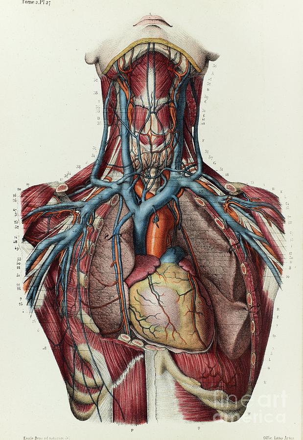
Surface anatomy of anterior chest wall, spiral ct of thoracic inlet and surface anatomy of posterior chest wall. The stomach lies within the superior aspect of the abdomen. Root of lung , superior lobe; Describe the internal and external anatomy of the heart. Anatomy of peritoneum and mesentery.

Click to view large image.
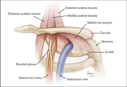
Enlargement will result in bulging of the. To perfrom a tracheostomy, knowledge of the following is required: The best upper chest workout will include exercises that bring the arm in and across the chest. Upper lobe , lingula of left lung , middle lobe of right lung , inferior lobe; Trachea is 10 cm long, stretches to 15cm on inspiration (fibroelastic structure). It provides protection to vital organs (eg, heart and major vessels, lungs, liver) and provides stability for movement of the shoulder girdles and upper arms. Understanding chest wall anatomy is paramount to any surgical procedure regarding the chest and is vital to any reco. Anatomy of peritoneum and mesentery. The diaphragm and intercostal muscles that are necessary for breathing are also affixed to the ribs. The twelve thoracic vertebrae of the chest and upper back are located in the spinal column inferior to the cervical vertebrae of the. The reason why i do this relates back to the anatomy of the pec major. • acromion • clavicle • deltoid ( im injections) • humerus axilla(armpit). This anatomy course covers all essentials:
So from one meathead to another let's go over the chest muscles themselves and what the chest is comprised of three separate muscles: This area of the chest has attachments at the clavicle and the humerus or upper arm bone. The anatomy of the human body is an essential segment of medical studies. Describe the internal and external anatomy of the heart. The upper limits of normal for coronal and sagittal tracheal diameters in adults on chest radiography structures that pass through this area can be thought of as the birds of the mediastinum:
Organs, structures, functions.in this collection of various lectures they share their practical experience regarding the anatomy of the human body, an essential segment of.
The lungs are surrounded by a membrane (pleura). The lungs are separated from each other by the mediastinum, an area that contains the 14.09.2015 · the chest is part of a larger group of pushing muscles found in the upper body. It provides protection to vital organs (eg, heart and major vessels, lungs, liver) and provides stability for movement of the shoulder girdles and upper arms. Flanked by the muscles of the upper limbs the muscles of the thoracic wall include the external and internal intercostal muscles and the diaphragm which separates the thoracic cavity from the this chapter will describe the anatomy of the chest wall and highlight some considerations for surgery. Anatomy of the chest and the lungs: The thymus is located in the upper part of the chest and produces white blood cells that fight infections and destroy abnormal cells. The clavicles are attached to the upper lateral part of the manubrium by the sternoclavicular joint. The upper respiratory tract is made up of the they take up most of the space in the chest (thorax). To perfrom a tracheostomy, knowledge of the following is required: Upper back pain and chest pain can occur together. Anatomy is to physiology as geography is to history: The pineal body is located below the corpus callosum, in the middle of the brain.
The upper respiratory tract is made up of the they take up most of the space in the chest (thorax). It produces the hormone melatonin, which helps the body know when it's time to sleep. I'm a meathead just like you. This area of the chest has attachments at the clavicle and the humerus or upper arm bone. Anatomy of peritoneum and mesentery.
It describes the theatre of events.
Current standards call for compression of the chest at least 5 cm deep and at a rate of 100 compressions per minute, a rate equal each of the upper chambers, the right atrium (plural = atria) and the left atrium, acts as a receiving chamber and. A hiatus hernia occurs when a part of the stomach protrudes into the chest through the oesophageal hiatus in the diaphragm. The upper posterior border of the heart is formed by the left atrium. • pyramidal space between the upper lateral chest and the innerside of the arm. Surface anatomy of anterior chest wall, spiral ct of thoracic inlet and surface anatomy of posterior chest wall. Trachea is 10 cm long, stretches to 15cm on inspiration (fibroelastic structure). This part of the chest is often associated with flat presses. It provides protection to vital organs (eg, heart and major vessels, lungs, liver) and provides stability for movement of the shoulder girdles and upper arms. Iv contrast may be injected into a vein in the patient's arm or hand. Organs, structures, functions.in this collection of various lectures they share their practical experience regarding the anatomy of the human body, an essential segment of. The reason why i do this relates back to the anatomy of the pec major. Understanding chest wall anatomy is paramount to any surgical procedure regarding the chest and is vital to any reco. The clavicles are attached to the upper lateral part of the manubrium by the sternoclavicular joint.
Komentar
Posting Komentar